|
© James H Nobbs |
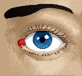
Sight can be considered as the most important of the senses, since it has been estimated that four-fifths of the information our senses gather about the world is through our eyes. The eyes transmit a constant stream of images to the brain by electrical signals. The eyes receive information from light rays. The light rays falling on most objects are either absorbed or reflected back. Objects that absorb all of the light rays appear black, whereas those that reflect all the light rays appear white. Coloured objects absorb certain parts of the light spectrum and reflect others. Colour, however, is not a property of the light, colour is a sensation perceived by the brain in a similar way to sound, touch, smell and taste.
In this section, the general functions of the eye are described first, and then the action of the light sensing cells is described in detail. The properties of the photoreceptors define the main features of our colour vision system.
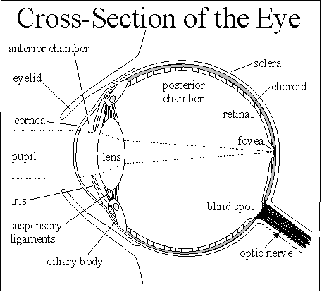
The internal structure of the eye is illustrated in Figure 2. When you look at something, the light rays reflected from the object enter the eye.
|
The light is refracted by the cornea and passes through the watery aqueous humor and pupil to the lens. The iris controls the size of the pupil and the amount of light entering the eye. The lens focuses the light which passes through the vitreous humor to form an image, reversed and upside down, on the retina.. Light-sensitive cells in the retina transmit the image to the brain by electrical signals. The brain "sees" the image right side up. |
|
The functions of the various structures of the eye can be understood by making a comparison with the four major functions of a camera
The internal structure of the eye is illustrated in Figure 2. The body of the eye is enclosed in a soft, flexible white casing known as the sclera, this is also known as "the white of the eye". A slight positive hydrostatic pressure inside the eye maintains a defined distance between the lens and the image plane (retina). The choroid is a brown, pigmented layer that absorbs stray light in the eye to reduce ghost images. The retinal layer is at the rear of the eye where the image is formed by the focusing system. The retina lies immediately above the choroid and is nourished through blood vessels in the choroid layer.
The cornea and the lens are the elements of the eye that focus the image onto the light sensitive area of the eye.
Light enters the eye through the cornea, a transparent section of the sclera that is kept moist and free from dust by the tear ducts and blinking of the eyelids. The shape of the cornea is determined by a slight positive hydrostatic pressure in the transparent liquid in chamber between the cornea and the lens (anterior chamber). The shape of the cornea is similar to that of a convex lens and it forms an image roughly in focus on the retina at the back of the eye. Too small a pressure in the liquid can cause far-sightedness. Too great a pressure in the liquid can cause nearsightedness.
The transparent flexible lens, whose shape is determined by radial muscles in the ciliary body, acts to form an inverted image of the field of view on the retina.
The distance from the lens to the retinal layer at the back of the eye is set by the vitreous humor, a viscose, transparent fluid present in the anterior chamber. This fluid is maintained at a slight positive pressure. A certain amount of waste tissue is suspended in the vitreous humor, this casts visible shadows on the retina when the image is a uniformly bright field such as the sky. The shadows are seen as nearly transparent strings of beads or intertwined ropes slowly moving around the field of view. The vitreous humor also contains very small particles in suspension that preferentially scatter blue light
Lens defects
The lens suffers from the two defects of chromatic and spherical aberration.
Chromatic aberration results in the blue and violet light being focused at a point closer to the lens than the green, yellow and red rays.
Spherical aberration results in the light rays passing through the outer edges of the lens being brought to focus at a different point to the rays passing through the centre of the lens.
The amount of light that passes into the eye is controlled by the diameter of the pupil. The pupil is formed by the iris, an annular shaped opaque layer whose inner diameter is controlled by the contraction and expansion of a set of circular and radial muscles.
The diameter of the pupil is small under high illumination (2 mm), but expands when illumination is low and may be as large as 8 mm for the dark-adapted eye. The colour of the iris may be black, brown, green or blue depending whether a lot, some, little or no brown pigment is present.
The retina is the light sensitive part of the eye and is made up of a mosaic of rod and cone shaped light sensitive cells, bipolar nerve cells and ganglions.
There are about 130 million sensory cells in the retina, of which 7 million are cone type cells. The rods and cones translate the optical image into a pattern of nerve activity that is transmitted to the brain by the million or so nerve fibres in the optic nerve.
Fovea
The image formed by the lens is centred on the fovea, an area of the retina that consists almost entirely of cone cells. This rod free region contains about 50,000 cone cells per square millimetre. It has the highest level of spatial resolution (visual acuity) of the image and the highest colour sensitivity. The fovea has a limited field of view, so the eyeball moves continuously to keep the image within it.
The nerve layer above the fovea is coloured with a yellow pigment and it is sometimes termed the yellow spot. The pigment serves to protect the fovea from over stimulation by filtering out some of the blue light. Normally this pigmented area is not apparent, as signals from the underlying cones have been adapted to the filtering effect. You may see it by placing a sheet of white paper before you and then closing the eyes for about 20 seconds. On opening the eyes, the projection of the yellow spot on the image of the white paper may be seen, however it will rapidly disappear as the retinal becomes adapted.
Blind spot
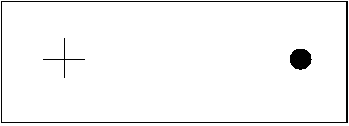
The blind spot is where the million or more nerve fibres that form the optic nerve leave the eye. This region contains very few light sensitive cells and the part of the image that falls on this area is invisible. Normally the gap in the visual field of one eye is filled by visual information from the other eye. The presence of the blind spot may be demonstrated with the aid of Figure 3.
Note that with one eye closed, it is normal to see no gap in the field of view; the missing area is filled in with an image of the same colour and texture as the surroundings. This adaptation must occur after the nerve signals have left the eye.
Light sensitive elements
|
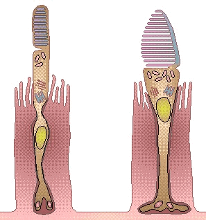
The shape and structure of the rod and the cone type cells are illustrated by Figure 4. The rods are about 1/100 of an inch (0.25 millimetres) long and about 1/400 of an inch (0.06 millimetres) thick. The cones are shorter and thicker. |
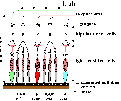
The light sensitive tips of the rods and cones are located deep within the retinal layer and lie next to the choroid coat of the eye, as shown in Figure 5.
|
The light must first pass through the nerve connections and blood vessels of the retinal layer before reaching the light sensitive region. The blood filled capillaries are semi-opaque, however the sensitivity of the underlying cones and rods adapt to this and the shadows cast are normally invisible. |
Figure 5: A cross section of the retina |
The cones and rods contain a photochemical pigment known as a photoreceptor, which is broken down and bleached by light. The photoreceptor pigment is present on the photoreceptor membrane, which looks like a stack of disks. A modified cilium connects the disks together. The pigment is synthesised in the inner segment of the rod or cone and is broken down and bleached by light. This breaking down process sets off an electrical charge that triggers the transmission of nervous impulses to the brain by way of the optic nerve. The interpretation of these impulses by the visual cortex to gives us the sensation of sight.
Rod cells
Only one type of rod shaped cell is present in the eye and they provide a monochrome light/dark view of the world. The sensitivity of rods to light depends on the presence of rhodopsin, a photosensitive pigment. Rhodopsin is continuously created in the eye and is destroyed by photo bleaching.
Bleaching is rapid in normal daylight, taking only a few minutes for completion. Under normal and high levels of illumination, only a small amount of rhodopsin is present and the rods have a low sensitivity too light.
When the illumination is reduced, the rate of bleaching is low and sensitivity slowly returns, taking up to thirty minutes to fully recover. At low levels of illumination (night or dark-adapted vision), the rods are sensitive to small amounts of light and are most sensitive to light with wavelengths close to 510 nm, a blue shade of green.
Cone cells
Three types of cone shaped cells have been identified in the eye with each type most sensitive to light of a particular range of wavelengths. The sensitivity of cones to light depends on the pigment iodopsin that is retained up to high levels of illumination. The combined responses of the three types of cone cell provide the ability to distinguish luminosity (light/dark) with the highest sensitivity at wavelengths near to 555 nm (green) of 683 lumens/Watt.
The three types of cone cell have been classified as long wavelength sensitive, medium wavelength sensitive and short wavelength sensitive corresponding to wavelength region of maximum response.
Short: Cones that are most sensitive to blue light, the maximum response being at a wavelength of about 440 nm (blue light).
Medium: Cones that are most sensitive to green light, the maximum in response being at a wavelength of about 545 nm (green light).
Long: Cones that are most sensitive to red light, the maximum in response being at a wavelength of about 585 nm (orange/red light).
Distribution of cone and rod cells
|
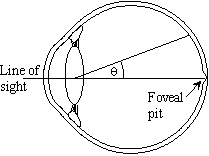
The distribution of cones and rods is not even across the retina. The distribution is best described in terms of areas contained within an angle θ to the line of sight drawn from the centre of the lens to the foveal pit as shown in Figure 6. |
|
The foveal pit occupies the central 2° field and is a rod free region that contains about 50,000 cones per sq. mm. An angle of 2° is presented by the image of a disc of diameter about 1 cm held 25 cm (the normal reading distance) from the eye.
The number of cones per unit area steadily decreases and the number of rods steadily increases in areas further out from the foveal pit
As the angle progressively increases beyond 20° the number of rods and cones per unit area steadily decreases.
There are only rods at angles greater than 40° so that peripheral vision is mainly monochromatic and is used mainly for the detection of movement.
Types of vision
Scotopic vision, low levels of illumination,
In dim light the rod cells are active and the cone cells are inactive. The rods cells provide black/white/grey vision only, which is called scotopic vision.
Photopic vision, medium and high levels of illumination
At medium and high illumination levels the cone cells are active and the rod cells are inactive. The three different types of cone cell provide colour vision, which is termed photopic vision.
Quality of vision
The fact that normal vision is clear, well defined and in colour is quite remarkable, since it is clear from this brief description that the signals provided by the eye are by no means perfect. The visual cortex has to correct for several defects.
The ability to correct for these defects is an illustration of the power of the visual cortex to process signals received from the optic nerve. This processing action has important effects on the way in which colour is sensed. It is not sufficient to just take into account the physiology of human eye; numerical colour description must also include the effects of this secondary visual processing.
|
© James H Nobbs |